‘Foamy’ microglia cells linked to more severe multiple sclerosis
21 May 2026
Multiple sclerosis (MS) is a chronic disease of the brain and spinal cord in which the immune system attacks myelin, the protective insulating layer surrounding nerve fibers. This demyelination can lead to neurological symptoms, including problems with walking, vision, and other neurological functions.
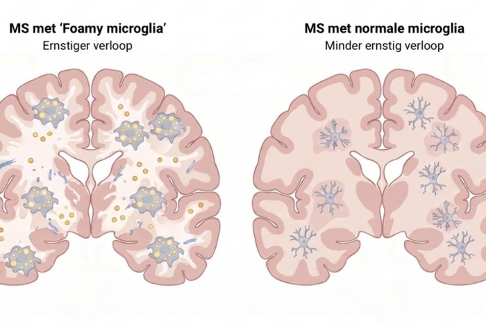
In a study just published in Nature Neuroscience, Daan van der Vliet and colleagues from the Netherlands Institute for Neuroscience, Leiden University and Utrecht University report a distinct population of lipid‑laden “foamy” microglia/macrophages in the brains of patients with secondary progressive multiple sclerosis (MS). Lesions enriched in these cells are closely associated with lesion expansion and a more severe disease course.
The lipids accumulating in these foamy microglia likely originate from damaged myelin that is being cleared by the cells. Under normal conditions, microglia help remove debris and damage in the brain, but in MS this task may become overwhelming. The researchers suggest that the cells take up such large amounts of damaged myelin that their waste-processing systems eventually become impaired, causing lipids to accumulate inside the cells.
These foamy lesions show disrupted lipid metabolism, lysosomal stress, and increased signatures of phagocytosis and antigen presentation yet lack classical pro‑inflammatory profiles. The team identifies the lipid‑metabolizing enzyme MAGL as a potential therapeutic target: its inhibition promoted lesion recovery in a mouse model of demyelination. Moreover, specific lipid species (oxylipins) in cerebrospinal fluid correlate with the proportion of foamy lesions, suggesting a promising route to fluid biomarkers for MS progression.